

Audio
Video
Transcript available via YouTube
Decode low neutrophils, recognize thalassemia without chasing iron, and sharpen your approach to bleeding disorders in primary care.
Crack the code on confusing labs. Learn from Dr. Bradley Beeler, an academic hematologist, how to recognize thalassemia without over-ordering tests, why a low neutrophil count in a healthy patient might not be a problem, and how to tackle bleeding disorders in primary care.
Claim CME for this episode at curbsiders.vcuhealth.org!
Patreon | Episodes | Subscribe | Spotify | YouTube | Newsletter | Contact | Swag! | CME
Show Segments
- Intro
- Case 1: Duffy-null associated neutrophil count
- Case 2: Bleeding disorders
- Case 3: Thalassemia
- Outro
Disclosures
Dr. Beeler reports no relevant financial disclosures. The Curbsiders report no relevant financial disclosures.
Pearls
- Avoid the term “benign ethnic neutropenia.” Replace it with Duffy-null–associated neutrophil count (DANC) to emphasize its genetic basis and reframe it as a normal physiologic variation.
- DANC is not pathologic. Most patients with Duffy-null–associated neutrophil count have no increased infection risk and do not need further workup if asymptomatic and stable.
- A neutrophil count of 1000–1500 can be normal, especially in individuals of African or Middle Eastern ancestry; further testing is often unnecessary unless ANC drops below 500 or symptoms develop.
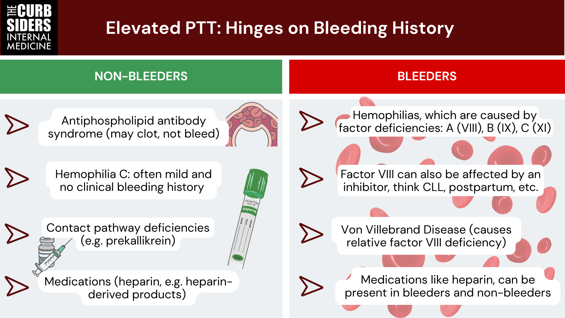
- Reframe elevated PTT by asking: Is the patient a bleeder? Use bleeding history to separate benign lab findings (e.g., contact pathway deficiencies) from true bleeding disorders (e.g., hemophilia, VWD).
- Von Willebrand disease does not protect against clots — Factor VIII replacement can overshoot and increase thrombotic risk; monitor levels in hospitalized patients receiving therapy.
- Mild thalassemia often masquerades as iron deficiency: Microcytosis with normal or elevated RBCs and iron studies should prompt evaluation for thalassemia, not empiric iron therapy.
- Many causes of elevated PTT don’t require treatment. Contact factor deficiencies or APS can prolong PTT without bleeding risk (APS carries a clotting risk, rather).
- Use ISTH-BAT to guide bleeding referrals: A score ≥4 in males or ≥6 in females supports evaluation by hematology.
- Don’t rely on hemoglobin electrophoresis to rule out alpha-thalassemia as it often misses mild disease; consider molecular testing if clinical suspicion remains high, but this testing is expensive and may be deferred for mild asymptomatic cases.
Benign Hematology Show Notes
Duffy-Null Associated Neutropenia (DANC)
Background
Duffy-null–associated neutrophil count (DANC) is caused by homozygosity for the rs2814778 single-nucleotide polymorphism (SNP) in the promoter region of the ACKR1 gene (formerly DARC), which encodes the Duffy antigen receptor for chemokines. This SNP disrupts a GATA1 transcription factor binding site, resulting in the absence of Duffy antigen expression on erythrocytes while preserving its expression on endothelium and other tissues. The Duffy-null phenotype is nearly fixed in many sub-Saharan African populations due to its protective effect against Plasmodium vivax malaria (Ferreira 2022), but it is also present at lower frequencies in Middle Eastern, Caribbean, Hispanic, Pacific Islander, and multi-racial populations (Bagheri 2022).
Prevalence
Greater than two-thirds or more of persons of African and Middle-Eastern descent can have this finding, and as a result have lower white blood cell counts (WBC) than patients of european descent (Bagheri 2022, Merz 2023).
Clinical Significance
There is no increased infectious risk in patients with DANC, and most patients can simply be reassured, especially if the finding is chronic and not associated with any symptoms. Always assess for symptoms in patients presenting with new neutropenia (expert opinion). Dr. Beeler likes to ask about infections, oral ulcers, fevers, and a broad review of systems is important to be sure a pathologic cause of neutropenia is not missed. Once it has been determined that the history and exam are most consistent with DANC, the patient can be reassured that it is likely a benign finding and does not require further evaluation (expert opinion).
Equity Implications
Dr. Beeler reminds the audience that DANC, a normal genetic variant most common among people of African and Middle Eastern ancestry, has too often been misclassified as pathologic, thus leading to unnecessary testing, referrals, and anxiety. Patients have been:
- Excluded from clinical trials because physiologic neutrophil ranges were misclassified as evidence of disease.
- Denied chemotherapy, as physiologic WBC counts are often interpreted as a treatment-limiting neutropenia.
- Subjected to unnecessary work-ups (e.g., bone-marrow biopsy) and the associated stress of believing they have a serious hematologic condition.
These harms reflect long-standing structural racism in lab reference ranges. To avoid pathologizing a normal trait, retire the term “benign ethnic neutropenia.” Instead, adopt the precise, person-centered term: Duffy-null–associated neutrophil count (DANC) (Hysong 2025, Merz 2023).
When to Refer to Hematology
Referral to hematology is appropriate when there are features concerning for an underlying hematologic disorder. These include constitutional symptoms such as weight loss, fevers, or night sweats; mucosal findings like oral ulcers; or a lack of historical support for DANC. For instance, if prior white blood cell counts were consistently in the normal range (expert opinion)
Dr. Beeler highlights that referral should also be considered when absolute neutrophil counts fall below 1000, and especially if they drop below 500, where DANC becomes less likely. That said, many individuals with DANC may have neutrophil counts in the 500–1000 range, and in asymptomatic patients with a stable, long-standing pattern of low neutrophils, further workup is often unnecessary (expert opinion, Merz 2023).
In ambiguous cases—especially when ANC is <1000—ACKR1 gene testing may be useful to confirm Duffy-null status. However, routinely sending genetic testing for all patients with ANC >1000 and a classic DANC phenotype may not represent high-value care. Testing should be targeted to cases where the diagnosis is unclear or where reassurance depends on objective confirmation (expert opinion, Ooga 2023).
Future Direction
In addition to replacing the term “benign ethnic neutropenia” with Duffy-null–associated neutrophil count, future efforts should focus on adopting ancestry-informed reference ranges that account for Duffy-null status, which may help reduce unnecessary testing and mitigate disparities in care (Merz 2022).
Bleeding Disorders Evaluation
Initial Framework
When abnormal bleeding is suspected based on history, primary care providers (PCPs) should be sure to elicit a detailed bleeding history by asking about the type and location of bleeding episodes, including mucocutaneous bleeding (such as epistaxis, gingival bleeding, easy bruising, or menorrhagia), deep tissue bleeding (such as hemarthrosis or muscle hematomas), and surgical or procedural bleeding. Clarifying these patterns can help differentiate between platelet-type disorders, von Willebrand disease, and coagulation factor deficiencies (Hughes 2024).
The ISTH Bleeding Assessment Tool (ISTH-BAT) can be used to quantify bleeding as normal or abnormal. Many of these questions are intuitive, and would likely be captured in a thorough history, but using the ISTH-BAT can help definitively outline abnormal bleeding and a need for further evaluation (Fasulo 2017). Notably, referral thresholds differ by sex: a BAT score of 4 or higher in males or 6 or higher in females should prompt consideration for referral to hematology.
The PCP can begin an initial workup which may include a complete blood count (CBC), prothrombin time and international normalized ratio (PT/INR), partial thromboplastin time (PTT), a basic chemistry panel, and iron studies to assess for potential anemia or underlying systemic causes (Hughes 2024, expert opinion).
Elevated PTT
When faced with an isolated PTT abnormality (a prolonged PTT with a normal PT/INR and no other obvious lab abnormalities), Dr. Beeler offers a practical diagnostic framework by dividing patients into two categories: “bleeders” and “non-bleeders.”
Bleeders
Among the “bleeders,” he includes conditions that are associated with both an elevated PTT and a bleeding tendency:
- Von Willebrand Disease (VWD): Typically a disorder of primary hemostasis, but VWD can also prolong the PTT because von Willebrand factor helps to prevent factor VIII from being broken down.
- Hemophilia A (Factor VIII deficiency): Caused by a deficiency of factor VIII and presents with bleeding into joints (hemarthroses), muscles, and after trauma or surgery.
- Factor VIII Inhibitor: This is an acquired condition in which autoantibodies inhibit factor VIII activity. It can present with severe, spontaneous bleeding in individuals without a personal or family history of hemophilia and often results in a dramatically prolonged PTT that does not correct with mixing studies. Often seen in postpartum females or patients with chronic lymphocytic leukemia (CLL) among other conditions.
- Hemophilia B (Christmas Disease): Similar in clinical presentation to Hemophilia A, but caused by a deficiency in factor IX. The PTT is prolonged, and bleeding may be spontaneous or triggered by minor trauma.
- Hemophilia C: A less common condition, usually seen in Ashkenazi Jewish populations, due to a deficiency in factor XI. It typically presents with mild to moderate bleeding, often post-operatively.
- Heparin or Other Drugs: Unfractionated heparin prolongs the PTT and may cause bleeding depending on the dose and context. Other anticoagulants (e.g., direct thrombin inhibitors like argatroban) may also contribute (Rountree 2023).
Non-Bleeders
When evaluating an isolated elevated PTT in a patient without any clinical signs of bleeding, Dr. Beeler suggests classifying them as a “non-bleeder.” These patients often have lab abnormalities that affect the intrinsic pathway but do not result in clinically significant bleeding. In fact, some may paradoxically be prothrombotic.
Common causes in non-bleeders include:
- Heparin exposure: Even small amounts of heparin or heparin-derived products can prolong the PTT.
- Hemophilia C (Factor XI deficiency): While technically a bleeding disorder, most patients are asymptomatic unless undergoing surgery. Routine bleeding is uncommon.
- Antiphospholipid syndrome (APS): A lupus anticoagulant can interfere with phospholipid-dependent clotting assays, leading to a prolonged PTT. However, these patients are more likely to clot than to bleed. A mixing study will fails to correct due to the presence of an inhibitor (antibody-mediated disease).
- Contact pathway deficiencies: These are rare and include prekallikrein, kallikrein, or high-molecular-weight kininogen deficiencies. Despite PTTs that may exceed 100 seconds, patients do not bleed. In these conditions. Prekallikrein is converted to kallikrein, which activates Factor XII, initiating the intrinsic cascade. A deficiency impairs lab-based clotting but not physiological hemostasis. A mixing study will correct.
Ultimately, if the patient has no bleeding history, a dramatically elevated PTT should be interpreted with caution. These “non-bleeders” often require no treatment, and in some cases (like APS), they may actually benefit from anticoagulation rather than hemostatic support.
Von Willebrand Disease
Types
Von Willebrand Disease (VWD) is the most common inherited bleeding disorder and is typically categorized into three main types. Type 1, which accounts for approximately 75–80% of cases, involves a partial quantitative deficiency of von Willebrand factor and follows an autosomal co-dominant inheritance pattern. Type 2 represents a qualitative defect, with several subtypes; notably, Type 2N (Normandy subtype) is characterized by defective binding to factor VIII, clinically mimicking hemophilia A: Dr. Beeler likes to remember this by there being “8” letters in “Normandy.” Type 3 VWD is the most severe form, marked by a near-complete absence of von Willebrand factor and therefore requiring prophylactic factor replacement therapy (expert opinion).
Management of VWD
One important pearl that Dr. Beeler mentioned is that VWD does not protect against thrombosis. In fact, some treatments, especially combination products containing both von Willebrand factor and factor VIII, can lead to supratherapeutic levels of factor VIII, increasing the risk of venous thromboembolism (VTE). Factor VIII levels should be monitored to remain within a safe therapeutic range (typically 50–150%), as levels above 150% are associated with increased thrombotic risk. For hospitalized patients receiving replacement therapy, daily inpatient monitoring is recommended. In the outpatient setting, if dosing is ongoing or clinical stability is uncertain, follow-up labs every 1–2 weeks are reasonable (Weyand 2021).
Finally, it is important to note that while VWD is classically associated with bleeding, some patients—particularly those with mild Type 1 or Type 2 disease—may not have overt bleeding symptoms and may fall into the “non-bleeder” category in certain contexts. However, this should not preclude appropriate perioperative or peripartum planning (expert opinion).
Postpartum Considerations
During pregnancy, von Willebrand factor and factor VIII levels naturally rise, but they may drop precipitously on postpartum days 3 to 4. This sudden decline can increase bleeding risk, particularly in the context of neuraxial anesthesia or surgical delivery, which can look like uterine atony but with an atypical timeline (VWF factors continue to drop up to three weeks from delivery). Therefore, close monitoring around the time of delivery is essential. Patients should be counseled on warning signs of postpartum hemorrhage and may benefit from antifibrinolytics like tranexamic acid or aminocaproic acid if bleeding develops (expert opinion).
Thalassemia
Microcytic Anemia Framework
Dr. Beeler uses the mnemonic “ISALT to recall the major causes of microcytic anemia. Here’s what it stands for:
- Iron deficiency anemia
- Sideroblastic anemia
- Anemia of chronic disease
- Lead poisoning
- Thalassemia
Thalassemia — General Principles
Thalassemia should be considered in patients who present with microcytic anemia, particularly when the RBC count is normal or elevated and iron studies are normal, indicating an iron-replete state. This pattern stands in contrast to iron deficiency, where the RBC count is typically low and ferritin levels are reduced (Baird 2022).
To confirm a suspected thalassemia, hemoglobin electrophoresis is the next step. In normal adults, hemoglobin composition is typically HbA ~97%, HbA2 ~2.5%, and HbF <1%. In beta-thalassemia trait, the hallmark finding is an elevated HbA2 level (>3.5%). In contrast, alpha-thalassemia is often missed on standard electrophoresis, as the hemoglobin fractions can appear normal unless the disease is more severe (e.g., HbH disease or Hb Barts in newborns). This distinction is rooted in the genetics: alpha-thalassemia involves two genes on chromosome 16, and deletions can be silent or subtle; beta-thalassemia is typically more detectable because of its effect on HbA2 levels (Baird 2022).
Thalassemia — Management
Management of thalassemia depends on the severity of the disease. For mild forms, treatment may include folic acid supplementation at a dose of 1 mg daily. In more severe cases, higher doses (2–5 mg/day) may be needed, though they can cause nausea. Importantly, iron supplementation should be avoided unless there is clear evidence of iron deficiency, as many thalassemia patients are iron-replete and at risk for overload (expert opinion).
For transfusion-dependent patients, management becomes more complex and includes regular phlebotomy or iron chelation therapy, along with osteoporosis screening and surveillance for endocrinopathies affecting the liver, heart, and gonads. Hepatitis A and B vaccination is also recommended due to increased transfusion-related infection risk (Borgna-Pignatti 2007, Baird 2022).
Clinically, many individuals with mild thalassemia have well-compensated anemia and may not require referral to hematology. However, genetic counseling becomes critical in prenatal care, especially for patients of Southeast Asian descent, where cis alpha-globin gene deletions can result in offspring at risk for hemoglobin Bart’s hydrops fetalis, a potentially fatal condition. Rather than the older terminology of “minor,” “intermedia,” and “major,” a more modern approach now distinguishes patients based on whether they are transfusion-independent or transfusion-dependent, which better reflects disease burden and management needs (Baird 2022, expert opinion).
Dr. Beeler’s CBC Clues That Warrant Hematology Referral
- Unexplained cytopenia
- Bicytopenia
- Blasts or dysplastic features on peripheral smear (e.g., hypogranularity, nuclear abnormalities)
- Elevated PTT with clinical bleeding
- Persistent neutropenia <1000, especially <500
Links
Goal
Listeners will explore how clinical pattern recognition and contextual clues can guide diagnosis in patients with common but frequently misunderstood blood disorders.
Learning objectives
- Recognize the clinical features and significance of Duffy-null–associated neutrophil count (DANC) and distinguish it from pathologic neutropenia.
- Apply a structured approach to evaluating bleeding symptoms using history, physical exam, and appropriate laboratory studies.
- Interpret an isolated elevated PTT by differentiating between bleeding and non-bleeding causes, including von Willebrand disease, hemophilia, and antiphospholipid syndrome.
- Identify hallmark clues for thalassemia on CBC and understand when to pursue further testing or hematology referral.
- Appreciate the equity implications of outdated reference ranges and adopt person-centered terminology and ancestry-informed interpretation of lab values.
Citation
Wurtz P, Beeler B, Williams PN, Watto MF. “491 Benign Hematology with Brad Beeler.” The Curbsiders Internal Medicine Podcast. thecurbsiders.com/category/curbsiders-podcast July 14, 2025.
Episode Sponsors
Panacea
Search The Podcast for Doctors (By Doctors) on Spotify, Apple Podcasts, or wherever you listen.
Locumstory
Locumstory.com is simply a free, unbiased educational resource about locum tenens.
Continuing Education Company
Curbsiders listeners get 45% off select online courses — that’s the biggest discount CEC has ever offered, and it’s exclusive to you with promo code Curb45, through July 30. You can also use Curb30 for 30% off all webcasts and on demand replay courses. Check it all out at CMEmeeting.org/curbsiders.
Episode Credits
Written and produced by Paul Wurtz MD. Show notes, cover art, and infographic also created by Paul Wurtz MD.
Hosts: Matthew Watto MD, FACP; Paul Williams MD, FACP
Reviewer: Sai S Achi MD, MBA, FACP
Showrunners: Matthew Watto MD, FACP; Paul Williams MD, FACP
Technical Production: PodPaste
Guest: Bradley Beeler MD
CME Partner

The Curbsiders are partnering with VCU Health Continuing Education to offer continuing education credits for physicians and other healthcare professionals. Visit curbsiders.vcuhealth.org and search for this episode to claim credit.
Contact Us
Got feedback? Suggest a Curbsiders topic. Recommend a guest. Tell us what you think.
We love hearing from you.
Join our Patreon!

Yes, you can now join our exclusive community of core faculty at Kashlak Memorial Hospital along with all the perks:
- Bundle & Save with the top CME tier!
- All episodes Ad-Free, including the entire back catalog!
- Twice-monthly bonus episodes: Ask me Anything (AMA) with Matt & Paul, Recaps, and more!
- Access to Kashlak's Discord Server!
- Advance Notice of Curbsiders Events!
- Exclusive Curbsiders' Merch!
