
Audio
Video
Transcript available via YouTube
A Bell-Ringer of a Presentation
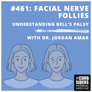
Is it a stroke or is it botox? No, it’s Bell’s palsy! Learn how to differentiate facial palsy from other sinister diagnoses. Join us as Dr. Jordan Amar (Assistant Professor of Clinical Neurology at USC Keck School of Medicine) gives us Bell’s palsy pointers (and a great mnemonic, if there is such a thing).
Claim CME for this episode at curbsiders.vcuhealth.org!
Patreon | Episodes | Subscribe | Spotify | YouTube | Newsletter | Contact | Swag! | CME
Show Segments
- Introduction
- Rapid fire questions and Reflex Hammers
- Case #1: Ms. Melanie
- Framework for Bell’s Palsy
- Bell’s Palsy vs Stroke: How to Differentiate
- Approach to History and Physical Exam
- Case #2: Mr. Lopez
- Red Flags in Facial Palsy (GROWLS FACE)
- Workup: From Imaging to Labs and What Not to Do
- Treatment: The Role of Steroids and Antivirals
- Supportive Care, Complications and Prognosis
- Take-Home Points
- Outro
Facial Nerve Follies: Understanding Bell’s palsy Pearls
- Bell’s palsy is defined as an isolated, spontaneous peripheral impairment of the 7th cranial nerve, with no other neurological or systemic symptoms. It typically presents with facial droop and forehead involvement, along with possible changes in taste, hyperacusis (increased hearing), and difficulty closing the affected eyelid.
- Bell’s palsy is a peripheral lesion that occurs after the nerve exits the brainstem (unlike central lesions, which occur at or above the brainstem).
- Unlike a stroke, which presents with sudden onset and fixed symptoms, Bell’s palsy builds gradually over 48-72 hours, reaching maximum severity within this time frame.
- Careful history and physical exam differentiate Bell’s palsy from other causes of facial palsy; ask about forehead involvement, hearing changes, recent infections (e.g., Lyme disease), and signs involving other cranial “neighborhood” nerves.
- The mainstay treatment is oral steroids (prednisone) initiated within 72 hours for maximum benefit. Antiviral therapy (e.g., valacyclovir) may be considered in certain high risk groups but is not strongly supported by evidence.
- Most patients with Bell’s palsy will see improvement within three weeks, with full recovery in 85% of cases within one year. Recurrence happens in about 7-8% of cases.
- Be wary of red flags! Worsening symptoms beyond 72 hours, presence of vertigo, hearing loss, bilateral facial palsy, involvement of other cranial nerves, or lack of improvement after three months are signs that something other than Bell’s palsy should be considered.
Facial Nerve Follies: Understanding Bell’s palsy –
Getting the Whole Story
Bell’s palsy is characterized by spontaneous peripheral impairment of the cranial nerve VII without other nerve involvement. Bell’s palsy is mostly considered idiopathic however, viral etiologies have been suspected (Baugh et al, 2013). The hallmark of a Bell’s palsy presentation is a combination of unilateral facial droop and blurred vision, which is distinct from other diagnoses on the differential. Forehead involvement, changes in taste, changes in hearing, and eyelid droopiness strongly point to peripheral VII facial nerve palsy (other diagnoses are still possible, however).
Consider cranial nerve VII impairment in patients with the following:
- Decreased taste in the anterior two-thirds of the tongue
- Hyperacusis due to loss of stapedius muscle function, making sounds louder – normal stapedius muscle functions to dampen sound
- Forehead involvement, which is present in Bell’s palsy – can be challenging to assess in Botox recipients (Tiemstra, 2007)
Additional historical symptoms that may point away from cranial nerve VII palsy include:
- Numbness, tingling of the face would suggest cranial nerve V palsy rather than Bell’s palsy
- Vision changes
- History of recent infections, tick exposure or rash (erythema chronicum migrans) that might suggest Lyme
- Autoimmune history, HIV infection or exposure
- Recent facial trauma (aka “Don’t forget the baseball bat!”)

It’s All About Timing
To help differentiate Bell’s palsy from a central lesion, ask the patient when they first noticed the symptoms, the speed of onset, and if the symptoms are worsening. Stroke symptoms typically present suddenly with maximal onset and are fixed, whereas Bell’s palsy often presents with a gradual build-up over 48 to 72 hours. Distinguishing between the two can be challenging, especially when patients wake up with symptoms. Bell’s palsy follows a monophasic course, where symptoms worsen, plateau, and do not worsen further after reaching the plateau.
Workup the VII
Classic idiopathic Bell’s palsy can be diagnosed through a thorough history and exam (Baugh et al, 2013). However, there should be a low threshold to pursue further workup if you spot red flag symptoms. Our expert suggests evaluating cranial nerve VII function with what he dubs the Jane Goodall Exam. Evaluate for asymmetry by asking the patient to smile and raise their eyebrows. Assess for facial muscle strength by tapping on the patient’s puffed out cheeks and attempting to open their tightly closed eyelids; ask the patient to close the eyelids “as though they had soap in them” and note for differences in strength. Time permitting, you could evaluate for loss of taste by applying a sugar packet to the anterior two-thirds of the tongue. Our expert reminds us that this could be difficult to execute, but it could be helpful.
Typical Physical Exam Findings in Bell’s palsy:
- A visible droop of the mouth on one side or a subtler flattening of the nasolabial fold
- Diminished wrinkling of the forehead on the affected side
- Lag or complete inability to close the eye on the affected side, with noticeable delay during blinking
- Inability to fully close the affected eye
- Increased sensitivity to sound (hyperacusis) on the affected side
- Diminished sweet taste sensation on the affected side of the tongue
On exam, be sure to examine the back of the mouth and the inner ear for any vesicles, lesions or other abnormalities. Look for “neighborhood signs,” which could suggest a brainstem lesion affecting cranial nerves V, VI, or VIII; these could manifest as changes in sensation, ocular movement, or hearing. Presence of these “neighborhood signs” argues AGAINST Bell’s palsy and FOR a brainstem lesion such as a stroke. To assess motor function, pay particular attention to arm weakness by examining for pronator drift (watch for pronation of the affected outstretched arm after asking the patient to hold out their arms as though holding a tray with closed eyes and while they shake their head). Another assessment Dr. Amar uses is the “orbiting test” wherein you have the patient to wrap their fists around each other. If one side is weaker, the stronger side will orbit around the weaker one, like a moon orbiting a planet.
It might not be Bell’s if…
The differential diagnosis for peripheral facial nerve palsy is extensive, including tumors, trauma, and various infections like Lyme disease, meningitis, HIV, tuberculosis and Ramsay-Hunt syndrome. Inflammatory conditions like sarcoidosis and neurological conditions like multiple sclerosis are also considerations. Guillain-Barre syndrome can cause bilateral Bell’s palsy BUT this is very rare, so Dr. Amar advises considering alternative processes alongside bilateral Bell’s palsy. More signs that point against Bell’s palsy include progression of symptoms beyond the previously noted typical 72-hour course and/or a lack of improvement at the 3-month mark point. Headache, dizziness and tinnitus are other symptoms that should point clinicians to an alternative diagnosis.

GROWLS FACE: Dr. Amar’s Trademark Mnemonic for Red Flags
Need a little help remembering red flags that point against Bell’s palsy? Our expert shared his mnemonic for red flags that may help (if you’re into mnemonics!): GROWLS FACE
- Gradual onset
- Risk factors for HIV
- Otitis media
- Worsening or no improvement within three months
- Limb involvement
- Systemic cancer
- Facial palsy with other symptoms
- Auditory canal or palate vesicles
- Cervical lymphadenopathy
- Ear pain or vesicles

Imaging, Labs and More
Dr. Amar tends to have a low threshold for imaging patients with findings that do not align with Bell’s palsy. While a standard brain MRI is sufficient, an internal auditory canal MRI is preferred for detailed visualization (Yücel et al., 2022). However, if stroke is suspected, the patient will require an immediate non-contrast CT to exclude bleeding. Laboratory testing should be guided by clinical suspicion for secondary causes such as Lyme disease, zoster and HIV testing. An electromyography (EMG) is not typically recommended due to discomfort and limited diagnostic value (expert opinion). However, EMG may be considered for prognostic information in long-term cases of Bell’s palsy (Guntinas-Lichius et al., 2020).
House Brackmann Scale
While many classification tools exist, the House Brackmann scale tends to be the most commonly used by neurology and ENT (House and Brackman, 1985). This scale is not mandatory for clinical practice but helps ENT assess the need for surgery and provides a baseline which can be then used to track response. This grading scale ranges from one to six, where one represents normal function and six represents total facial paralysis. The scale allows subjective findings to be quantified for both neurologists and primary care physicians and is a useful before-and-after measure for treatment. One drawback is that the inter-user reliability is questionable. In his practice, Dr. Amar takes photos of the patient and puts the photos in the patient’s medical record to monitor disease progression and treatment response.
Treatment
Idiopathic Bell’s palsy
Treatment typically involves oral steroids such as prednisone (Gronseth and Paduga, 2012; Jalali et al, 2021). Our expert recommends starting prednisone 60-80 mg daily for 5 days and tapering off; some clinicians may choose to taper the dose after the initial five days, but this remains an expert opinion. For better outcomes, Dr. Amar reminds us to start treatment as early as possible, particularly within 72 hours of symptom onset. In his personal practice, he does start treatment as late as one week from symptom onset in people who are immunocompromised.
Evidence supporting antiviral treatment is limited, and its effectiveness relies on concurrent steroid use. Antiviral agents like acyclovir or valacyclovir may be added if herpes involvement is suspected. In more severe cases, especially if the patient is at risk for a herpes zoster virus infection, both steroids and antivirals may be considered, depending on the patient’s ability to tolerate both medications.
In summary, our expert finds that if the patient has severe Bell’s palsy, and has low risk for side effects from antivirals, the potential benefit may outweigh the low risk of short term antiviral treatment once other potential diagnoses have been ruled out.

Supportive Measures
Eye Care
As previously discussed, the recovery process for patients with Bell’s palsy can be slow and full of lingering issues. In Dr. Amar’s expert opinion, the primary goal is to lower the risk of exposure keratopathy (drying and subsequent ulceration of the cornea due to excessive exposure to the open air from an incompletely closing eyelid). Lubricating eye drops, ointments and eye patches are standard recommendations; other methods, such as taping the eye shut, are also appropriate so long as the eye is closed and covered (Baugh et al, 2013). In Dr. Amar’s experience, eye patches are not typically preferred by patients so, the compromise is limiting the usage of the eye patch to sleep, as that is a period where damage to the cornea is at high risk. A rare surgical solution for patients with eyelid weakness refractory to treatment is the implantation of small weights in the affected eyelid (Rahman and Sadiq, 2007). These eyelid weight procedures are largely cosmetic and are unlikely to offer superior protection of the eye when compared to an eye patch or tape per Dr. Amar.
Pain Management
Bell’s palsy can lead to chronic neuropathic pain, which is often difficult to manage. Despite the limited evidence supporting their efficacy, Dr. Amar’s expert opinion and clinical experience suggest that antiepileptics such as carbamazepine and phenytoin could be therapeutic options. These medications also tend to cause less sedation compared to other common treatments like gabapentin.
Facial Physical Therapy
A small recent study tried a combination of passive and active stretching of facial muscles as well as electrically stimulating the facial nerve to ameliorate the effects of Bell’s palsy and showed significant improvement in its participants (Santiago et al., 2024). While trials such as this one are small and have not yet been widely reproduced, facial physical therapy can help physical improvement and recovery from a long-term, potentially traumatizing condition.
Long Term Sequelae
Chronic residual Bell’s palsy can result in a phenomenon called synkinesis, due to compensatory innervation of the muscles around those that were affected by their palsy. In synkinesis, you will see activation of muscles that would typically not be activated in stereotyped patterns of movements (Guntinas-Lichius et al., 2022).
Another long-term sequelae of Bell’s palsy is lacrimal gland changes. Initially, loss of lacrimal gland function will cause drying of the eyes, but long term, there will be a rebound increase in tearing that will occur during actions such as talking or laughing, causing patients to cry what are known as “crocodile tears” (Modi and Arsiwalla, 2023).
Prognosis and Recovery
Dr. Amar encourages us to reassure our patients that many improve within the first three weeks. By the three to six-month mark, around 75 percent will have significant improvement (Baugh et al, 2013).
Take-Home points
- Bell’s Palsy is isolated, peripheral involvement of the CN VII that gradually peaks in 48-72 hours
- Be cognizant of the red flags, aka: GROWLS FACE
- Ask about trauma, aka “Don’t Miss the Bat” — Dr. Amar
Links


Goal
Listeners will develop a medical care strategy for Bell’s palsy from diagnosis to treatment
Learning Objectives
After listening to this episode listeners will…
- Recognize the defining characteristics and diagnostic criteria of Bell’s palsy
- Formulate an approach to diagnose and differentiate Bell’s palsy from other conditions
- Develop a plan for the medical management of Bell’s palsy in a timely manner
- Establish proactive measures to prevent complications in patients with Bell’s palsy
Disclosures
Dr. Jordan Amar and The Curbsiders report no relevant financial disclosures.
Citation
Valdez I, Jordan A, Williams PN, Watto MF. “#461: Facial Nerve Follies:
Understanding Bell’s Palsy”. The Curbsiders Internal Medicine Podcast. thecurbsiders.com/category/curbsiders-podcast Nov 11, 2024.
Episode Credits
Producer and Writer: Isabel Valdez, PA-C
Show Notes: Paul Wurtz MD, Ben Furman MPH MS3, Isabel Valdez, PA-C
Infographic and Cover Art: Zoya Surani, MS2
Hosts: Matthew Watto MD, FACP; Paul Williams MD, FACP
Reviewer: Leah Witt, MD
Showrunners: Matthew Watto MD, FACP; Paul Williams MD, FACP
Technical Production: PodPaste
Guest: Jordan Amar, MD
CME Partner

The Curbsiders are partnering with VCU Health Continuing Education to offer continuing education credits for physicians and other healthcare professionals. Visit curbsiders.vcuhealth.org and search for this episode to claim credit.
Contact Us
Got feedback? Suggest a Curbsiders topic. Recommend a guest. Tell us what you think.
We love hearing from you.
Join our Patreon!

Yes, you can now join our exclusive community of core faculty at Kashlak Memorial Hospital along with all the perks:
- Bundle & Save with the top CME tier!
- All episodes Ad-Free, including the entire back catalog!
- Twice-monthly bonus episodes: Ask me Anything (AMA) with Matt & Paul, Recaps, and more!
- Access to Kashlak's Discord Server!
- Advance Notice of Curbsiders Events!
- Exclusive Curbsiders' Merch!
